hisology course كورسات تعليمية لمادة الهستولوجي

الموضوع في 'الفيديوهات والبرامج الطبية' بواسطة Dr.Sayed, بتاريخ ‏ديسمبر 8, 2005.

  1. Dr.Sayed

    Dr.Sayed مشرف سابق

    إنضم إلينا في:
    ‏مارس 18, 2005
    المشاركات:
    611
    الإعجابات المتلقاة:
    175
    نقاط الجوائز:
    0
    الوظيفة:
    طالب بالفرقة الثالثة بكلية الطب البيطري - جامعة ال
    مكان الإقامة:
    جمهورية مصر العربية


    بسم الله الرحمن الرحيم

    الأخوة والأخوات ..

    أقدم لكم إن شاء الله سلسلة تعليمية شاملة لكل فروع مادة الهستولوجي أو علم الأنسجة والدورة إن شاء الله مدعمة بالصور والشرح المختصر وبالإختبارات والأسئلة على كل فرع من فروعها .... والدورة مقدمة لكل طلاب كليات الطب بجميع فروعها وطلاب اقسام البيولوجي بكليات العلوم والتربية وكل من يدرس هذه المادة المهمة وأرجو أن تفيدكم وأدعو الله عز وجل أن يعينني على إكمالها وإخراجها بالشكل الائق...

    والسلام عليكم ورحمة الله وبركاته
     
    nonameforever و rana elshabasy معجبون بهذا.
  2. jam1966

    jam1966 مشرف عام سابق

    إنضم إلينا في:
    ‏نوفمبر 3, 2004
    المشاركات:
    3,734
    الإعجابات المتلقاة:
    3,802
    نقاط الجوائز:
    128
    الجنس:
    ذكر
    مكان الإقامة:
    بلاد العرب اوطاني
    وفقك الله ورعاك اخي د سيد

    تحياتي وتقديري لك
     
  3. Dr.Sayed

    Dr.Sayed مشرف سابق

    إنضم إلينا في:
    ‏مارس 18, 2005
    المشاركات:
    611
    الإعجابات المتلقاة:
    175
    نقاط الجوائز:
    0
    الوظيفة:
    طالب بالفرقة الثالثة بكلية الطب البيطري - جامعة ال
    مكان الإقامة:
    جمهورية مصر العربية
    Epithelia

    Epithelia
    Epithelia are tissues consisting of closely apposed cells without intervening intercellular substances. Epithelia are avascular, but all epithelia "grow" on an underlying layer of vascular connective tissue. The connective tissue and the epithelium are separated by a basement membrane. Epithelium covers all free surfaces of the body. Epithelium also lines the large internal body cavities, where it is termed mesothelium. Furthermore, the internal surfaces of blood and lymph vessels are lined by epithelium, here called endothelium.
    Epithelia are classified on the basis of the number of cell layers and the shape of the cells in the surface layer.
    *If there is only one layer of cells in the epithelium, it is designated simple.
    *If there are two or more layers of cells, it is termed stratified.
    *Cells in the surface layer are, as a rule, described according to their height as squamous (scale- or plate-like), cuboidal or columnar.
    =====================================================================
    1-Simple Epithelia
    a-Simple squamous epithelium
    This type is composed of a single layer of flattened, scale- or plate-like cells. It is quite common in the body. The large body cavities and heart, blood vessels and lymph vessels are typically lined by a simple squamous epithelium. The nuclei of the epithelial cells are often flattened or ovoid, i.e. egg-shaped, and they are located close to the centre of the cells.
    [​IMG]

    b-Simple cuboidal epithelium
    Cells appear cuboidal in sections perpendicular to the surface of the epithelium. Viewed from the surface of the epithelium they look rather like small polygons. Simple cuboidal epithelium occurs in small excretory ducts of many glands, the follicles of the thyroid gland, the tubules of the kidney and on the surface of the ovaries.
    [​IMG]

    c-Simple columnar epithelium
    The cells forming a simple columnar epithelium are taller than they are wide. The nuclei of cells within the epithelium are usually located at the same height within the cells - often close to the base of the cells. An example is the simple columnar epithelium which lines the internal surface of the gastrointestinal tract (GIT) from the cardia of the stomach to the rectum.
    [​IMG]

    Note:- Identifying Epithelia?
    The outlines of individual epithelial cells are not always visible, and it may be difficult to identify the shape of the cells. It is often helpful to look at the shape, location and spacing of the nuclei in the epithelium, which together will allow a very good guess at the shape of the cells forming the epithelium.
    [​IMG]

    =======================================================================
    2-Stratified Epithelia
    a-Stratified squamous epithelium
    Stratified squamous epithelia vary in thickness depending on the number of cell layers present. The deepest cells, which are in contact with the basement membrane, are cuboidal or columnar in shape. This layer is usually named the basal cell layer, and the cells are called basal cells. Basal cells are mitotically active and replace the cells of the epithelium which are lost by "wear and tear". The basal cell layer is followed by layers of cells with polyhedral outlines. Close to the surface of the epithelium, cells become more flattened. At the surface of the epithelium, cells appear like flat scales - similar to the epithelial cells of simple squamous epithelia.
    Remember that it is the shape of the cell which form the surface of the epithelium which gives the name to the epithelium!
    [​IMG]

    b-Stratified cuboidal and columnar epithelia
    are not common. A two-layered cuboidal epithelium is, for example, seen in the ducts of the sweat glands. Stratified columnar epithelia are found in the excretory ducts of the mammary gland and the main excretory duct of the large salivary glands.
    [​IMG]


    Stratified columnar epithelia
    [​IMG]


    Stratified cuboidal epithelia
    [​IMG]

    ================================================================
    3-Pseudostratified and Transitional Epithelia
    These two types of epithelia are difficult to classify using the shape of the cells in the surface layer and the number of the cell layers as criteria.
    a-Transitional epithelium
    Transitional epithelium is found exclusively in the excretory urinary passages (the renal calyces and pelvis, the ureter, the urinary bladder, and part of the urethra).
    The shape of the cells in the surface layer of a transitional epithelium varies with the degree of distension of the organs whose lumen is lined by this type of epithelium. In the "relaxed" state of the epithelium, it seems to be formed by many cell layers. The most basal cells have a cuboidal or columnar shape. There are several layers of polyhedral cells, and, finally, a layer of superficial cells, which have a convex, dome-shaped luminal surface. In the distended state of the epithelium only one or two layers of cuboidal cells are followed by a superficial layer of large, low cuboidal or squamous cells. In the distended state the epithelium will resemble a stratified squamous epithelium.
    [​IMG]

    b-Pseudostratified columnar epithelium
    All cells of this type of epithelium are in contact with the basement membrane, but not all of them reach the surface of the epithelium. Nuclei of the epithelial cells are typically located in the widest part of the cell. Consequently, the nuclei of cells which do or do not reach the surface of the epithelium are often located at different heights within the epithelium and give the epithelium a stratified appearance. The epithelium will look stratified but it is not - hence its name "pseudostratified". Pseudostratified columnar epithelia are found in the excretory ducts of many glands.
    [​IMG]


    [​IMG]

    ======================================================================​
     
  4. Khaled_Hadhoud

    Khaled_Hadhoud New Member

    إنضم إلينا في:
    ‏أكتوبر 9, 2005
    المشاركات:
    415
    الإعجابات المتلقاة:
    118
    نقاط الجوائز:
    0
    الوظيفة:
    DOCTOR
    مكان الإقامة:
    EGYPT
    شكرا جزيلا لك أخى د. سيد

    وفقك الله
     
  5. Dr.Sayed

    Dr.Sayed مشرف سابق

    إنضم إلينا في:
    ‏مارس 18, 2005
    المشاركات:
    611
    الإعجابات المتلقاة:
    175
    نقاط الجوائز:
    0
    الوظيفة:
    طالب بالفرقة الثالثة بكلية الطب البيطري - جامعة ال
    مكان الإقامة:
    جمهورية مصر العربية
    شكراً

    شكراً أخي الدكتور خالد وأخي جام على المرور والرد.:clap: :clap: :heart:
     
  6. Dr.Sayed

    Dr.Sayed مشرف سابق

    إنضم إلينا في:
    ‏مارس 18, 2005
    المشاركات:
    611
    الإعجابات المتلقاة:
    175
    نقاط الجوائز:
    0
    الوظيفة:
    طالب بالفرقة الثالثة بكلية الطب البيطري - جامعة ال
    مكان الإقامة:
    جمهورية مصر العربية
    Glands

    GLANDS
    are cells or aggregations of cells whose function is secretion.
    *Exocrine glands: release the secretory product via a system of ducts that opens upon one of the surfaces of the body which are in contact with the external world (skin, gastrointestinal tract etc.).
    *Endocrine glands: release their secretory product (typically hormones) into the spaces between the secretory cells (extracellular space) from which it enters the bloodstream.
    Both endocrine and exocrine glands are developmentally derived from epithelia, which form a down-growth into the underlying connective tissue. The cells forming this down-growth then develop the special characteristics of the mature gland. Exocrine glands maintain the connection with the body surface whereas it is lost by endocrine glands.
    --------------------------------------------------------------------------------
    *Classification of Exocrine Glands:
    Exocrine glands may be classified according to cell number, and/or the shape and branching pattern of their secretory portions and ducts.
    *Unicellular Glands
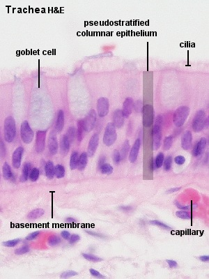
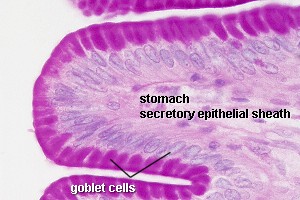
    consist of a single secretory cell. In mammals the only example of unicellular exocrine glands are goblet cells, which occur in the epithelium of many mucous membranes. Goblet cells secrete the glycoprotein mucin, which by the uptake of water is converted into a slimy substance, mucus.
    [​IMG]
    unicellular exocrine glands (goblet cells)
    *Multicellular glands :
    The simplest form of a multicellular gland is a secretory epithelial sheath - a surface epithelium consisting entirely of secretory cells (e.g. the epithelium lining the inner surface of the stomach, where the mucous secretion protects the stomach wall from the acidic contents of the stomach). Other multicellular glands have their secretory portion embedded in the connective tissue underlying the epithelium. The secretion is either discharged directly from the secretory portion onto the epithelium or reaches the epithelium via a duct system that consists of non-secretory cells.
    [​IMG]
    The secretory portion may have a variety of shapes. Secretory cells may form
    *tubes in tubular glands,
    *acini in acinar glands or
    *alveoli in alveolar glands
    [​IMG]
    Combinations exist - the pancreas is a tubulo-acinar gland, in which each section of the secretory system has a specialized function.
    The precursors of digestive enzymes are produced by the acinar cells. Tubular cells secrete the alkaline bicarbonate solution which eventually neutralizes the acidic contents of the stomach that are released into the duodenum.
    Multicellular glands with an unbranched excretory duct are called simple. We talk about a compound gland when the excretory duct is branched. Finally, the part of the gland consisting of secretory cells is branched in a (surprise!) branched gland.
    The classification scheme may appear somewhat elaborate - but there are many exocrine glands around. All of them can be identified and described by this scheme, and some ideas about their function can be derived from this description.
    Secretory Mechanisms
    The secretory cells can release their secretory products by one of three mechanisms.
    1.Merocrine secretion corresponds to the process of exocytosis. Vesicles open onto the surface of the cell, and the secretory product is discharged from the cell without any further loss of cell substance.
    [​IMG]
    2.Apocrine secretion designates a mechanism in which part of the apical cytoplasm of the cells is lost together with the secretory product. The continuity of the plasma membrane is restored by the fusion of the broken edges of the membrane, and the cell is able to accumulate the secretory product anew. This mechanism is used by apocrine sweat glands, the mammary glands and the prostate.
    [​IMG]
    3.Holocrine secretion designates the breakdown and discharge of the entire secretory cell. It is only seen in the sebaceous glands of the skin.
    [​IMG]
    There are two additional mechanisms by which secretory cells can release their products. Lipid soluble substances may diffuse out of the secretory cell (e.g. steroid hormone producing endocrine cells). Transporters (membrane proteins) may actively move the secretory product across the plasma membrane (e.g. the acid producing parietal cells of the gastric glands). These secretory mechanisms may not involve any light microscopically visible specialisations of the cell.
    Histological Structure of Large Exocrine Glands:
    The relationship between the secretory tissue (parenchyma) of glands and the supporting connective tissue is similar in most larger glands. Externally the entire gland is surrounded by a layer of dense connective tissue, the capsule. Connective tissue sheets (septa) extend from the capsule into the secretory tissue and subdivide the gland into a number of lobes. Thinner connective tissue septa subdivide the lobes into a number of lobules. Reticular connective tissue (hardly visible in H&E stained sections) surrounds and supports the secretory units of the glands (alveoli, acini etc.) and the initial parts of the excretory ducts if present.
    Blood and lymph vessels as well as nerves penetrate the capsule and form a delicate network between the secretory units and the initial parts of the duct system.
    The main excretory duct conveys the secretory product to one of the external surfaces of the body. Other parts of the duct system are named according to their relation to the lobes and lobules of the gland.
    *Lobar ducts are are large branches of the main duct which extend to the lobes of the gland. They may be called Interlobar ducts.
    *Interlobar ducts if they are found in the connective tissue surrounding the lobes. Interlobar ducts branch and give rise to Interlobular ducts.
    *Interlobular ducts, which are found in the connective tissue surrounding the individual lobules of the gland. Branches of the interlobular ducts enter the lobules and are now called Intralobular ducts.
    *Intralobular ducts. The terminal branches of the duct system, which connect intralobular ducts with the secretory units of the gland, are called Intercalated ducts.
    *Intercalated ducts.
    The appearance of the different portions of the duct system is quite variable from gland to gland and may allow the identification of the gland. Quite often, the appearance of parts of the duct system also permits some deductions about their functions.
    *Note that lobes and lobules are defined by their relationship to each other. Many small lobules may form one large lobe. Neither size nor the spatial relationship between different parts of the tissue can be unequivocally determined in a single, two-dimensional section of the tissue. Lobes and interlobar ducts may therefore be difficult to distinguish from lobules and interlobular ducts.
    [​IMG]
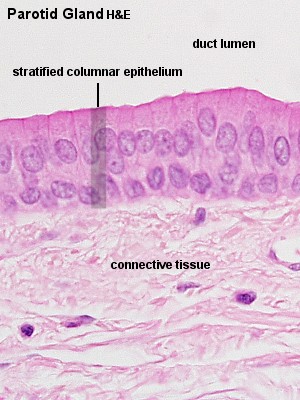
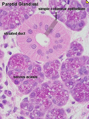
    Parotid Gland:alveolar gland
    [​IMG]
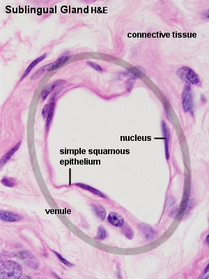
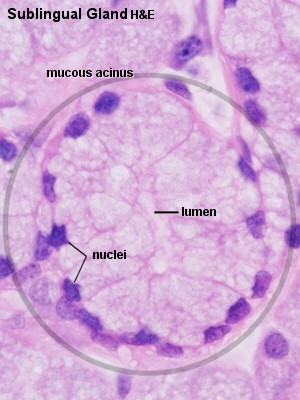
    Sublingual Gland:alveolar gland

    ==============================================================​
     
  7. sini

    sini Guest

    شكرا لكم على هذا الموقع الرائع و الله يجازيكم الخير
     
    sini,
  8. Dr.Sayed

    Dr.Sayed مشرف سابق

    إنضم إلينا في:
    ‏مارس 18, 2005
    المشاركات:
    611
    الإعجابات المتلقاة:
    175
    نقاط الجوائز:
    0
    الوظيفة:
    طالب بالفرقة الثالثة بكلية الطب البيطري - جامعة ال
    مكان الإقامة:
    جمهورية مصر العربية
    تم رفع الصور الى مركز تحميل الموقع وجاري اعداد الدرس الجديد إن شاء الله.
     
  9. mehdi

    mehdi New Member

    إنضم إلينا في:
    ‏سبتمبر 21, 2005
    المشاركات:
    117
    الإعجابات المتلقاة:
    0
    نقاط الجوائز:
    0
    بارك الله فيك اخي الكريم Dr.Said على الموضوع الرائع
     
  10. فاطمةالجندي

    فاطمةالجندي New Member

    إنضم إلينا في:
    ‏ديسمبر 2, 2009
    المشاركات:
    4
    الإعجابات المتلقاة:
    0
    نقاط الجوائز:
    0
    جزاكم الله خيرا
     

مشاركة هذه الصفحة